.

My life, my beat.

Dank der Kardiologie Basel sportlich unterwegs

Cardiology

The heart sets the rhythm. If you are worried about your heart, we are here for you. At our practice Herz-Lungen-Praxis, a team of specialists awaits you. They will give you and your health their full attention. and use state-of-the-art technology for support during diagnosis and treatment. With us, you will always speak to the same trustworthy doctor. We respond to your needs and concerns and respect your desire for privacy.

Prevention is key

A personal check-up provides you with preventive information about your state of health. Early detection and treatment of high blood pressure, for example, is an important preventive measure.

If other diagnostic measures are necessary – such as a cardiac catheter, CT scan or MRI – you and the referring doctor will make arrangements for these to be scheduled at a hospital of your choice. We maintain good relationships with all the surrounding hospitals.

We take time to listen

The patient consultation is central to our work. We respond thoroughly to your questions and concerns. Since information about pre-existing medical conditions also helps us make a diagnosis, please bring existing medical documents with you to your appointment. You are welcome to bring a companion to accompany you. We will find the path that is right for you. It is important to us that you are actively involved in making decisions about your care.

Women and men with the same illness often display different symptoms. We take this into consideration. We can perform urgent exams at short notice, and we provide continuous care for chronically ill patients. It's your health, after all.

Tests

To conduct a 24-hour blood pressure measurement, a blood pressure monitor is worn for 24 hours. This allows blood pressure to be assessed over a longer period of time than a single measurement. It also allows the dip in blood pressure that normally occurs at night to be assessed.

24-hour blood pressure measurement can provide information about

  • High and low blood pressure
  • Presence or absence of the nighttime dip in blood pressure
  • Need for treatment of previously undetected high blood pressure
  • Treatment monitoring of high blood pressure that is already being treated

Procedure

The patient receives a small device to take home. This device is worn with a belt and an arm cuff. The patient should go about their normal daily routine. The device cannot be worn in the shower or bath. Activities, symptoms and sleep times should be recorded in the report provided. Once the device is returned, the readings are analyzed and the findings are discussed with the patient.

Duration

Approx. 15 minutes to attach the device

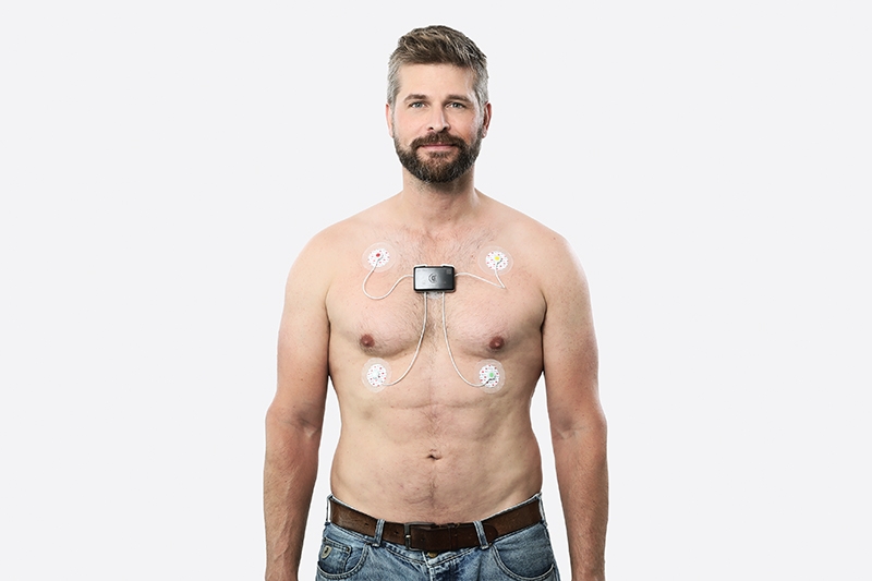
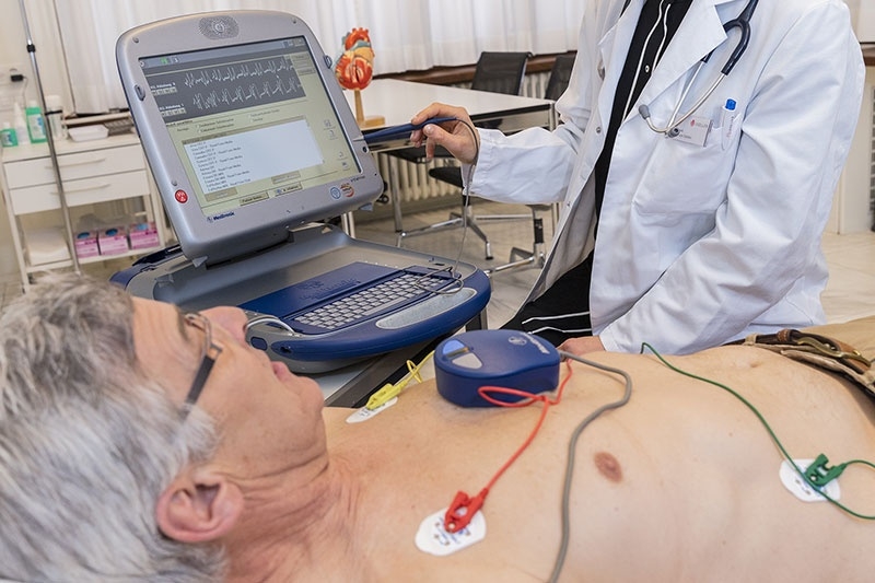
A 24-hour ECG takes an ECG over an entire day. It allows the cardiac rhythm to be assessed over a longer period of time than a resting ECG.

It can provide information about

  • Irregular heartbeat (occurring frequently)
  • Causes of syncope (loss of consciousness)
  • Causes of tachycardia, skipped heart beat, palpitations, vertigo
  • Monitoring of treatment of arrhythmia with medication

Procedure

The patient receives a small device to take home. This device is worn with a belt. The electrodes are attached to the chest with adhesive. The patient should go about their normal daily routine. The device cannot be worn in the shower or bath. Symptoms and sleep times should be recorded in the report provided. Once the device is returned, the data is analyzed. The findings are discussed with the patient.

Duration

Approx. 15 minutes to attach the device

24-h EKG_am Model_1_bearb

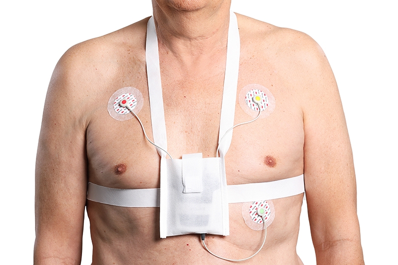
A 7-day ECG records an ECG over an entire week. It allows the cardiac rhythm to be assessed over a longer period of time than a 24-hour ECG.

It can provide information about

  • Irregular heartbeat (occurring infrequently)
  • Causes of syncope (loss of consciousness)
  • Causes of tachycardia, skipped heart beat, palpitations, vertigo
  • Monitoring of treatment of arrhythmia with medication

Procedure

The patient receives a small device to take home. This device is worn with a belt. The electrodes are attached to the chest with adhesive. The patient should go about their normal weekly routine. The patient may shower or bath during the 7-day ECG. To do so, the device must be removed. After showering, new electrodes are attached. Symptoms and sleep times should be recorded in the report provided. Once the device is returned, the data is analyzed. The findings are discussed with the patient.

Duration

Approx. 15 minutes to attach the device

7-Tage EKG_am Model_2_bearb

An abdominal ultrasound allows various organs within the abdomen to be examined, such as the liver, kidneys, pancreas and spleen. It is a non-invasive, painless examination.

Procedure

The patient lies on their back.

On the morning of the exam, the patient should not eat or drink, since the contents of the intestines could block the view.

Duration

Approx. 30 minutes

Viollier_Abdomen_Echo_001

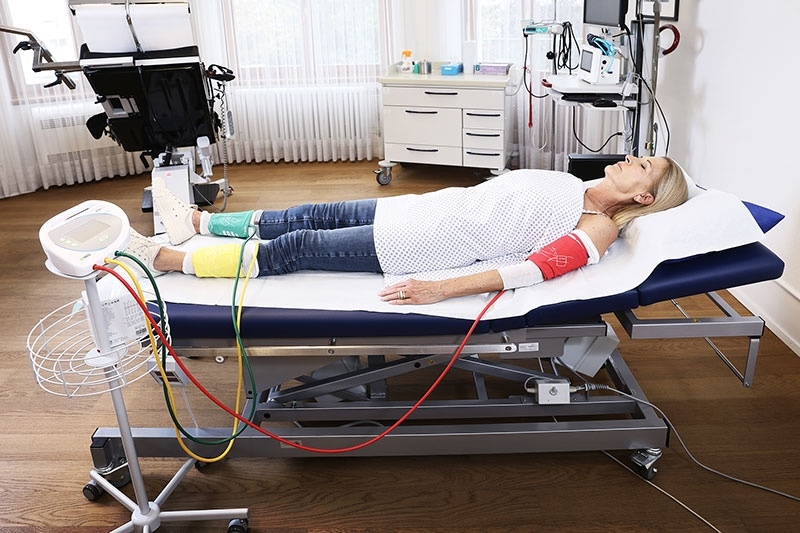
The ankle-brachial index is a simple method of detecting peripheral arterial disease. It is a good predictor of later cardiovascular complications.

It can provide information about

  • The presence of peripheral arterial disease (PAD) at an ABI of < 0.9, even if the patient is asymptomatic.
  • Occurrence of later cardiovascular complications at an ABI of < 0.9.

Procedure

The patient lies on their back. After a short period of rest, blood pressure is measured at the upper arm and at the ankle.

Duration

Approx. 20 minutes including resting phase

Kardiologie Basel Ankle-Brachial-Index (ABI)

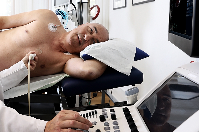
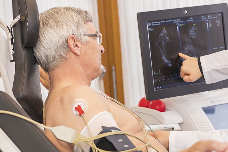
The cardiac ultrasound is a non-invasive test that allows the heart to be viewed in detail without radiation. This test allows the structure of the heart (wall thickness, heart valves) and the blood flow through the heart to be examined more closely by means of ultrasound.

Scope of application

  • Change in the heart at high blood pressure
  • Investigation of heart murmur
  • Monitoring after a heart attack
  • Myocarditis or pericarditis
  • Explanation of shortness of breath
  • Explanation of hereditary heart conditions

It can provide information about

  • Heart valve structure (leaky valves, constrictions)
  • Heart muscle structure (thickening, thinning)
  • Heart muscle function (pumping function)
  • Examination of large blood vessels

Procedure

The patient lies on their left side. The left chest area is examined with an ultrasound probe. An ECG is taken at the same time.

Duration

Approx. 1 hour for consultation with the doctor, preparation, Resting ECG, echocardiogram

Viollier_Herz_Echo_001

Dynamic stress echocardiography is used to test the heart under stress to provide evidence of possible circulatory disorders. In contrast to stress echocardiography with dobutamine, the patient's heart rate is increased by physical exertion on a stationary bicycle, and no medication is given. This test is only possible if good resting ultrasound images can be generated and if the patient can handle sufficient exertion on the bicycle.

It can provide information about

  • Circulatory disorders of the heart under stress
  • Changes in blood flow due to valve changes under stress

Procedure

The patient lies on a special recumbent bicycle. An ECG is performed during physical exertion. The intensity increases continuously over the course of the test. If symptoms such as chest pain, shortness of breath or exhaustion occur, the test is terminated.

Comfortable clothing or athletic clothing for cycling is ideal.

Duration

Approx. 2 hours for consultation with the doctor, preparation, infusion, Resting ECG, cycling

KAR_Dynam_Stressecho_19

An implantable event recorder allows the rhythm of the heart to be recorded over several years. A small device is implanted directly beneath the skin and measures cardiac current. If arrhythmia occurs, a signal is sent automatically to the attending physician's computer. The doctor can then view the ECG online. If the patient notices symptoms beginning, they can also send a signal and the doctor can view the ECG from this period of time in detail.

It can provide information about

  • Infrequent arrhythmia that cannot be detected with the 7-day ECG
  • Causes of syncope (loss of consciousness)
  • Causes of infrequently occurring symptoms such as tachycardia, skipped heart beat, palpitations

Procedure

The device is implanted here at the practice. The patient receives a local anesthetic.  The procedure usually involves one or two stitches so that the incision heals quickly. The stitches can be removed at the heart and lung practice or by the patient's general physician after 7 days. The device is then checked every 6 months.

In the event of symptoms, the patient can generate a signal.

If the device records a relevant irregularity in heart rhythm, the doctor receives a message.

Duration

Approx. 1.5 hours onsultation with the doctor, preparation, implantation

In certain types of arrhythmia, implanting a pacemaker may be necessary. For severe irregularities in heart function, some patients receive an implantable defibrillator (ICD). Implantation under the skin of the chest takes place in a hospital. The devices are regularly read at our practice to test their functionality and adjust the settings if needed. The device is tested to make sure it is working and the battery level is checked. Possible cardiac irregularities that have been recorded in the device's memory are read.

Procedure

The pacemaker and ICD device is checked every 6 – 12 months. The patient is lying down during the test. A scanning head is placed on the device from the outside. The doctor discusses with the patient whether the device's functions need to be adjusted to meet the patient's needs.

Duration

Approx. 1 hour for consultation with the doctor, preparation, Resting ECG, pacemaker scan

Kardiologie Basel Herzschrittmacher- und ICD-Kontrolle

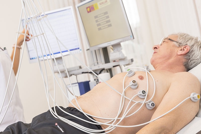
The resting ECG records a weak current from the heart.

It can provide information about

  • Arrhythmia and heart impairment
  • Abnormal changes in heart structure
  • Circulatory disorders of the heart

Procedure

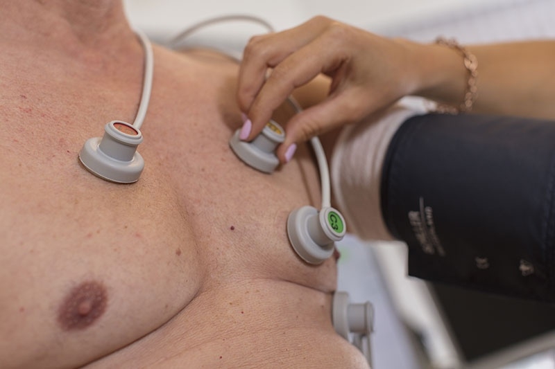
The patient lies resting for several minutes while an ECG is performed with a total of 12 electrodes.

Duration

Approx. 15 minutes including preparation

Kardiologie Basel Ruhe-EKG (Elektrokardiogramm)

A stress ECG (ergometry) involves an ECG being taken while the patient is subjected to increasing exertion on a bicycle. Blood pressure is also measured at regular intervals.

It can provide information about

  • Individual resilience
  • Impaired circulation in coronary heart disease
  • Hypertension under stress
  • Irregular heartbeat under stress

Procedure

The patient sits on a stationary bicycle. An ECG is taken while the patient cycles. Blood pressure is also measured. The intensity increases continuously over the course of the test. If symptoms such as chest pain, shortness of breath or exhaustion occur, the test is terminated.

Comfortable clothing or athletic clothing for cycling is ideal.

Certain medications should be stopped before the stress test. Our team will gladly provide you with the appropriate information.

Duration

Approx. 1 consultation with the doctor, preparation, resting ECG, cycling

Kardiologie Basel Belastungs-EKG (Ergometrie)

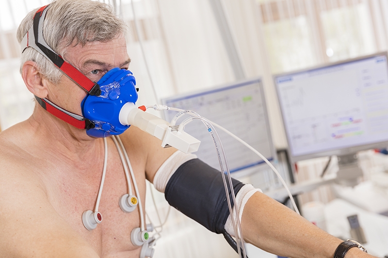
Spiroergometry consists of a stress ECG conducted while wearing a special mask. The mask is used to examine oxygen absorption by the tissue, the anaerobic threshold (exertion limit) and breathing under stress.

It can provide information about

  • Causes of shortness of breath
  • Evaluation of patients with cardiac insufficiency
  • Evaluation of athletes' performance
  • Exertion recommendations for patients after rehabilitation

Procedure

The patient sits on a stationary bicycle. An ECG is taken while the patient cycles. Blood pressure is also measured. The respiratory mask measures the respiratory rate, the respiratory volume, and levels of oxygen and carbon dioxide in the exhaled air. The intensity increases continuously over the course of the test. If symptoms such as chest pain, shortness of breath or exhaustion occur, the test is terminated.

Comfortable clothing or athletic clothing for cycling is ideal.

Certain medications should be stopped before spiroergometry. Our team will gladly provide you with the appropriate information.

Duration

Approx. 1.5 hours for consultation with the doctor, preparation, Resting ECG, cycling

KAR_Spiroergometrie_03

A stress echocardiogram is performed in the diagnosis of circulatory disorders. Unlike the resting ECG, here the heart is examined under stress. The patient receives a medication that causes the heart rate to increase. This allows any circulatory disorders of the heart to be confirmed, since these more frequently occur under stress when the heart muscle receives too little oxygen.

It can provide information about

  • Circulatory disorders of the heart under stress
  • Changes in blood flow due to valve changes under stress

Procedure

As with an echocardiogram, the patient lies resting on their left side. The patient also receives a cannula before the exam, through which the medication to increase the heart rate is given.

Certain medications should be stopped before this examination. This is discussed during the pre-procedure consultation with the patient. A consent form must be signed in advance.

Duration

Approx. 2 hours for preparation and consultation with the doctor, stress echocardiogram

In certain situations, an ultrasound of the heart must be performed from the esophagus. This provides clearer images than those produced from outside the body.

It can provide information about

  • Thromboses (blood clots) in the heart
  • Inflammation of the heart valves (endocarditis)
  • The condition of the heart valves for planning of heart valve surgery
  • Congenital defect joining the right and left ventricles and the atria
  • Examination of the aorta (main artery)

Procedure

The patient lies down and an intravenous line is placed in the arm. The throat is numbed with a spray. The ultrasound probe is inserted into the esophagus, similar to a gastroscopy.

The patient must fast (no food) for 8 hours before the procedure.

Duration

Approx. 1 hours for preparation and consultation with the doctor, transesophageal ultrasound

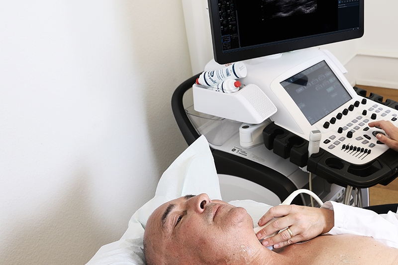
This ultrasound can assess the blood vessels going to and from the heart and those leading to the brain.

It can provide information about the blood vessels such as

  • Changes in the vascular wall
  • Plaque buildup
  • Constriction
  • Dilation

Procedure

The patient lies on their back and is examined with an ultrasound probe.

A vascular ultrasound is usually performed after an echocardiogram.

Duration

Approx. 10 minutes

Viollier_Gefaess_Echo_001

Pneumology

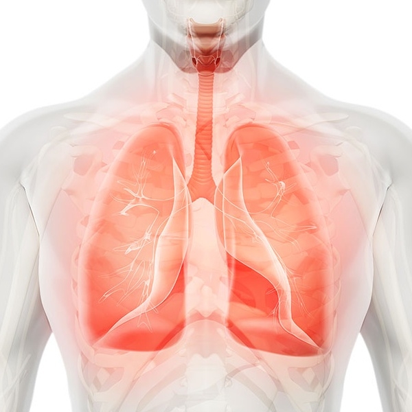
The lungs provide us with the oxygen we need to survive. If oxygen absorption is impaired, shortness of breath seriously limits quality of life. Our pulmonologists are here for you and will help you catch your breath so you can achieve better quality of life.

More

Pneumologie Basel bei Lungenbescherden

Herz-Lungen-Praxis Spalenring 147
4055 Basel

T +41 848 121 121
Contact

Opening hours Monday – Friday
07:30 – 18:00 h

Arrival Public transport
Bus no. 50
Tram no. 1, no. 6 Brausebad

Swiss Accreditation SMTS 0069
Swiss Accreditation